Home » Without Label » Anatomy Muscles Pelvis / MRI pelvis anatomy | free male pelvis axial anatomy ... - Psoas consists of a pair of deep muscles (psoas major and iliacus) located on each side of the pelvis in the abdomen.
Anatomy Muscles Pelvis / MRI pelvis anatomy | free male pelvis axial anatomy ... - Psoas consists of a pair of deep muscles (psoas major and iliacus) located on each side of the pelvis in the abdomen.
Anatomy Muscles Pelvis / MRI pelvis anatomy | free male pelvis axial anatomy ... - Psoas consists of a pair of deep muscles (psoas major and iliacus) located on each side of the pelvis in the abdomen.. Comparatively, there is much more movement at the pectoral girdle than at the pelvic girdle. See more ideas about anatomy, thoracic, basic image. The floor of the pelvis is made up of the muscles of the pelvis, which support its. On the posterior side they are the glutei and on the anterior side the hip muscles extending into the thighs. The levator ani is a broad sheet of muscle.
It takes origin from the inner aspect of pelvis along a line extending from the body of the pubis to the ischial spine. Describe the muscles of pelvic diaphragm. The inferior aspect of the pelvic cavity is called the pelvic diaphragm. Enclosing and protecting abdominopelvic and pelvic viscera. Look at the pictures and appreciate how complex the anatomy is.
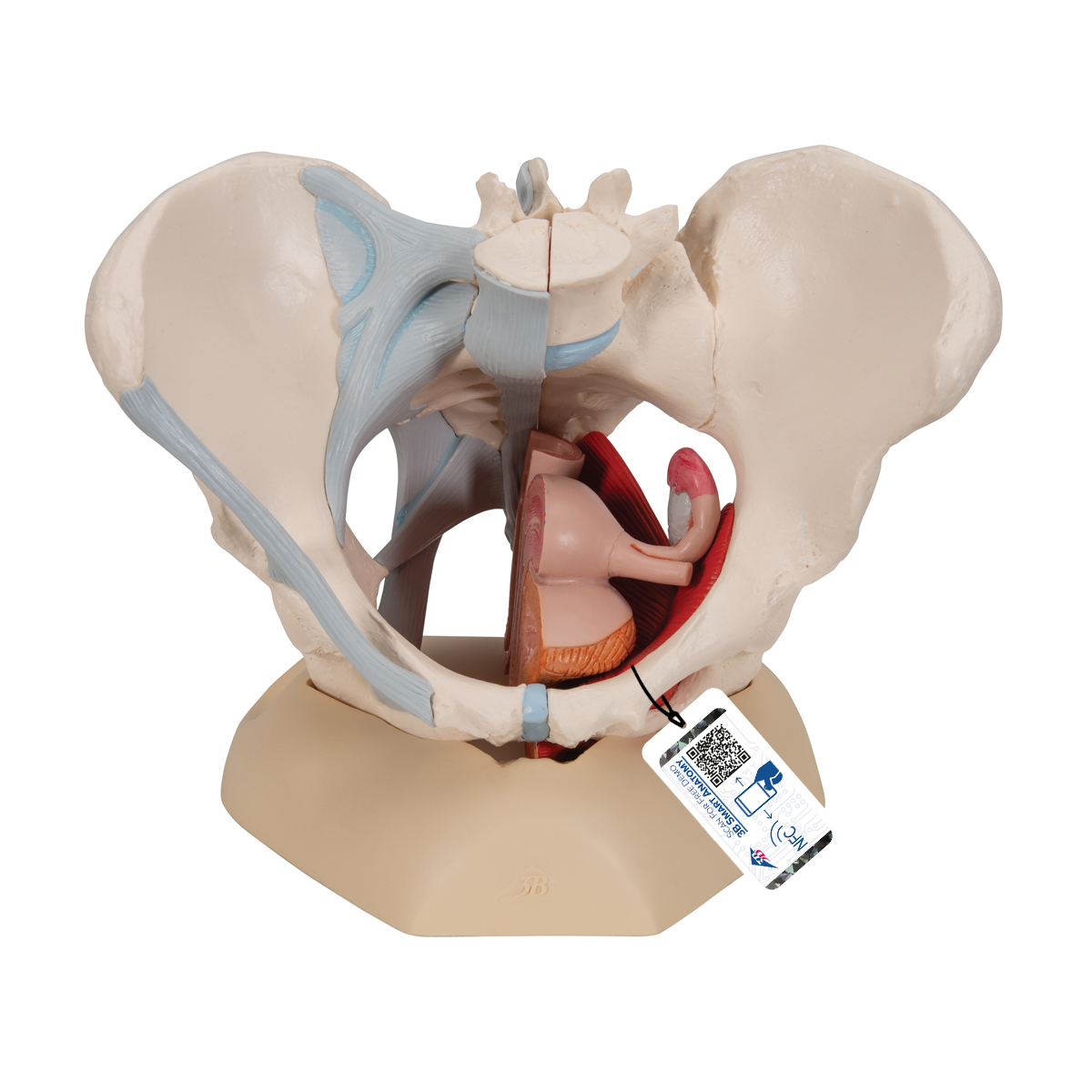
Anatomical Teaching Models - Plastic Human Pelvic Models ... from www.3bscientific.com In this video, we explore the anatomy of the pelvic diaphragm muscles of the pelvic floor, The hip bone attaches the lower limb to the axial skeleton through its articulation with the sacrum. These muscles have attachments to the pelvis as follows: They form a large sheet of skeletal muscle that is thicker in some areas than in others. The medial surface provides attachment for both transverse perinei, obturator internus and externus, piriformis, coccygeus and levator ani muscles. To maintain the continence of urine and faeces. The pelvic floor muscles include; The levator ani is a broad sheet of muscle.
The floor of the pelvis is made up of the muscles of the pelvis, which support its.
Muscles that attach from the pelvis to the trunk and cross the lumbosacral joint muscles that attach from the pelvis to the thigh/leg and cross the hip joint pelvic floor muscles that are located wholly within the pelvis There is very little movement of the pelvic girdle because of its connection with the sacrum at the base of the axial. The pelvic cavity and perineum. The medial surface provides attachment for both transverse perinei, obturator internus and externus, piriformis, coccygeus and levator ani muscles. The right and left hip bones, plus the sacrum and the coccyx, together form the pelvis. The pelvic girdle is formed by a single hip bone. Included in this group are the adductor longus, adductor brevis, adductor magnus, pectineus, and gracilis muscles. To support the abdominal and pelvic viscera. It takes origin from the inner aspect of pelvis along a line extending from the body of the pubis to the ischial spine. The pelvic floor muscles include; The inferior aspect of the pelvic cavity is called the pelvic diaphragm. Look at the pictures and appreciate how complex the anatomy is. On the posterior side they are the glutei and on the anterior side the hip muscles extending into the thighs.
Area between the asis (anterior superior iliac spine) and aiis (anterior inferior iliac spine). False pelvis is the pelvis major and is the space between the iliac crests (iliac fossae) its inferior boundary is the pelvic brim. This mri male pelvis axial cross sectional anatomy tool is absolutely free to use. It works better during single movements. The main function of the pelvic floor muscles are:
Pelvic Floor Muscles Anatomy Ct | Review Home Co from i1.wp.com Describe the muscles of pelvic diaphragm. To support the abdominal and pelvic viscera. Attached to the pelvis are muscles of the buttocks, the lower back, and the thighs. To maintain the continence of urine and faeces. The floor of the pelvis is formed by the two muscles named levator ani and coccygeus. The floor of the pelvis is made up of the muscles of the pelvis, which support its. The muscles that make up the pelvis diaphragm are the piriformis, coccygeus, iliococcygeus, pubococcygeus, and puborectalis. Anterior part of the medial condyle of the tibia.
The muscles of the pelvis and hip control the vast range of movement of the legs and torso.
It works better during single movements. The muscles that make up the pelvis diaphragm are the piriformis, coccygeus, iliococcygeus, pubococcygeus, and puborectalis. Helps improve kegel and pelvic floor. These muscles, including the gluteus maximus and the hamstrings, extend the thigh at the hip in support of the body's weight and propulsion. Attached to the pelvis are muscles of the buttocks, the lower back, and the thighs. False pelvis is the pelvis major and is the space between the iliac crests (iliac fossae) its inferior boundary is the pelvic brim. A proper kegel exercise means a full contraction and relaxation of the pc muscle. Muscles an important group of muscles in the pelvis is the pelvic floor. The pelvis's frame is made up of the bones of the pelvis, which connect the axial skeleton to the femurs, and therefore acts in weight bearing of the upper body. Providing attachment for many muscles and ligaments used in locomotion; To support the abdominal and pelvic viscera. The pelvis is the lower portion of the trunk, located between the abdomen and the lower limbs. Area between the asis (anterior superior iliac spine) and aiis (anterior inferior iliac spine).
The levator ani muscles are the largest group of muscles in the pelvis. The ischium provides numerous points of attachment for pelvic and lower limb muscles. Don't worry about the details. As well as some basic images of disc pathology and stylised facet joint motion. The muscles that make up the pelvis diaphragm are the piriformis, coccygeus, iliococcygeus, pubococcygeus, and puborectalis.
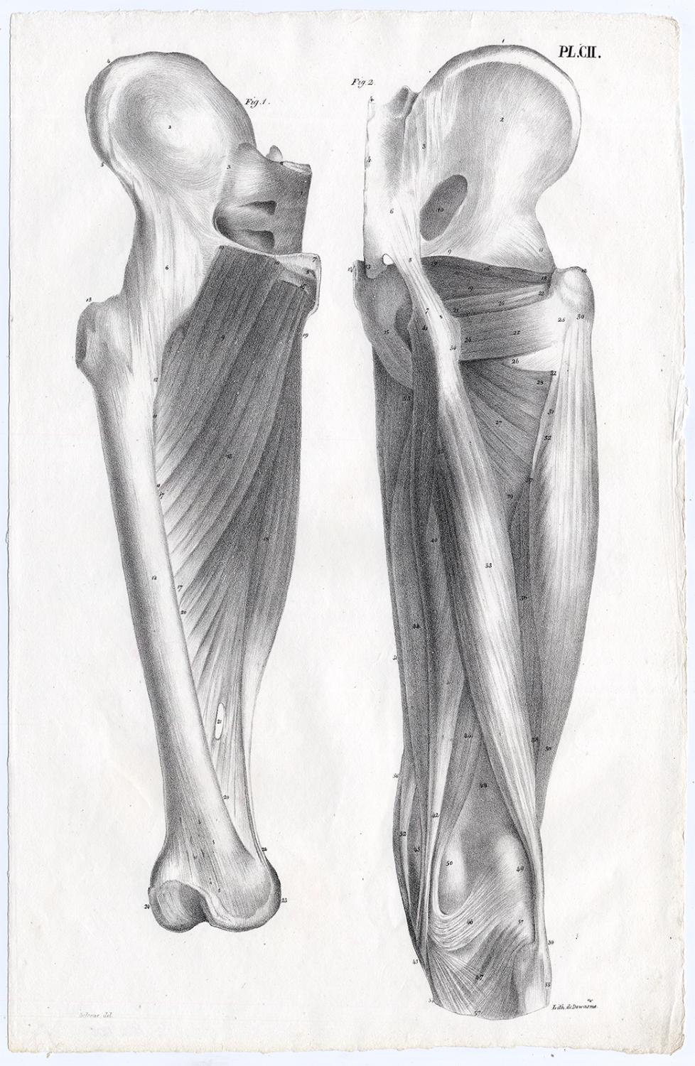
Antique Print-HUMAN ANATOMY-MUSCLES-LEG-FEMUR-BONY PELVIS ... from pictures.abebooks.com The floor of the pelvis is made up of the muscles of the pelvis, which support its. True pelvis is the pelvis minor and is the space inferior to the pelvic brim. It is a broad flat muscle. In this video, we explore the anatomy of the pelvic diaphragm muscles of the pelvic floor, The pelvic cavity and perineum. The inferior aspect of the pelvic cavity is called the pelvic diaphragm. The pelvis is the lower portion of the trunk, located between the abdomen and the lower limbs. They have several functions, including helping to support the pelvic organs.
The muscles of the pelvic floor are collectively referred to as the levator ani and coccygeus muscles.
Currently most of the pictures are of the female pelvis, i will add the male pelvis when i have time. These muscles move the thigh toward the body's midline. See more ideas about anatomy, thoracic, basic image. The iliacus muscle is part of a complex muscle system in the hip area that can function on its own or with other muscles. Describe the muscles of pelvic diaphragm. The pelvis's frame is made up of the bones of the pelvis, which connect the axial skeleton to the femurs, and therefore acts in weight bearing of the upper body. It separates pelvis from the perineum. Comparatively, there is much more movement at the pectoral girdle than at the pelvic girdle. The pelvis is the lower portion of the trunk, located between the abdomen and the lower limbs. It can be described as one of the bodies diaphragms. The pubococcygeus (pc) muscle is the muscle that runs the show in pelvic floor health. They have several functions, including helping to support the pelvic organs. There is very little movement of the pelvic girdle because of its connection with the sacrum at the base of the axial.